Visit ParagonCare Indonesia
 Visit ParagonCare Japan
 Visit ParagonCare New Zealand
 Visit ParagonCare Hong Kong
 Visit ParagonCare Indonesia
 Visit ParagonCare Korea
 Visit ParagonCare Malaysia
 Visit ParagonCare Singapore
 Visit ParagonCare Thailand
 Visit ParagonCare Vietnam


Nidek
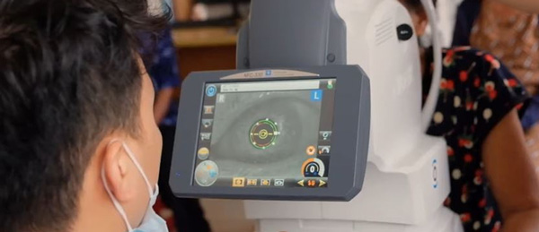
NIDEK is committed to promoting continuous technological innovation to develop solutions that meet the evolving needs of eye care professionals, supporting both the Quality of Vision (QOV) and Quality of Life (QOL) of patients worldwide.
Optical Coherence Tomography RS-1 Glauvas
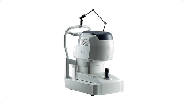
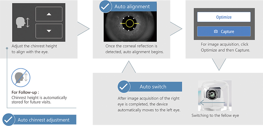
Effortless operation and interpretation
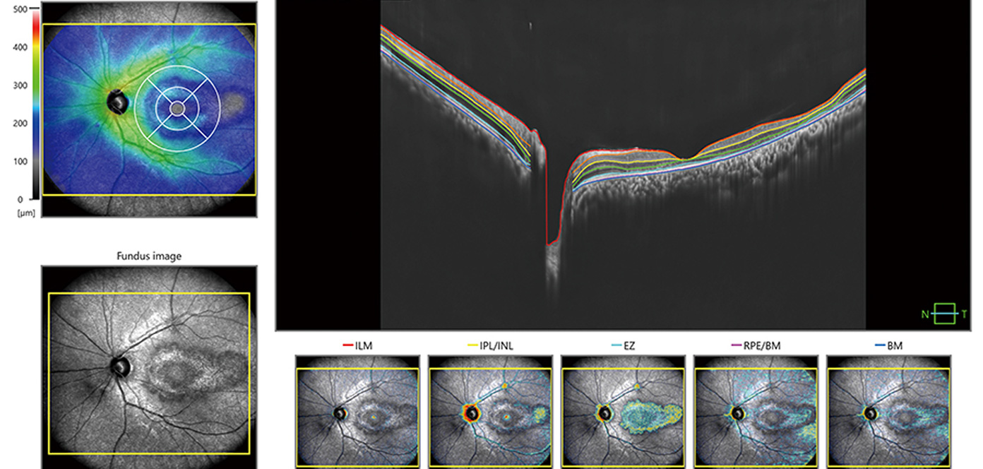
The RS-1 Glauvas delivers high-speed, high-resolution OCT imaging, capturing 250,000 A-scans per second for wide, deep and detailed views of ocular structures. Designed for effortless operation and interpretation, it pairs advanced analytics with an intuitive workflow to support faster, more confident clinical decision-making.
Advanced analytics
The incorporation of 250,000 A-scans/s accelerates your workflow by reducing capture time. The high-speed imaging also addresses patient fixation errors thus contributing to greater image clarity and patient comfort.
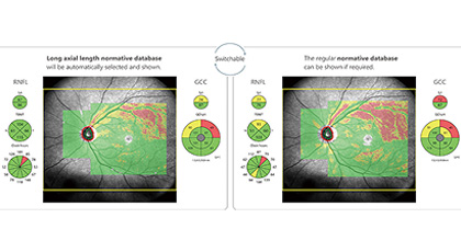
Glaucoma analysis in myopia
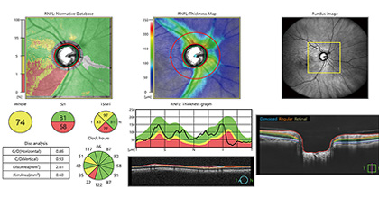
Less false positives
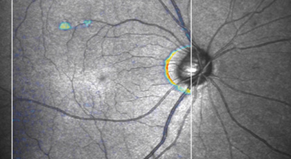
Structural Normality Map(SN Map) improving diagnostic confidence on early detection
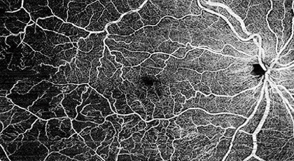
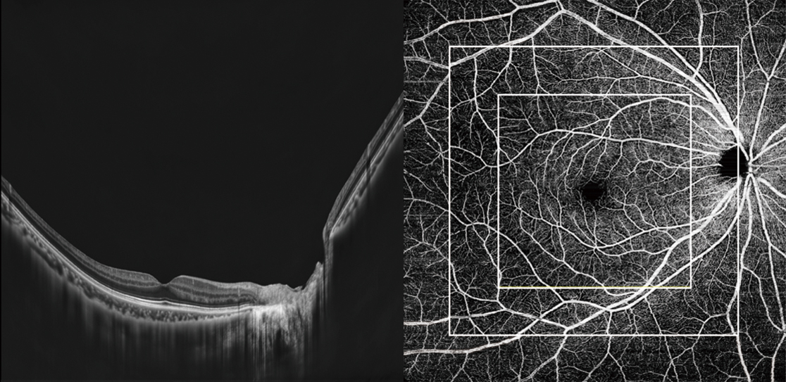
Enhanced OCT-A resolution with a new imaging algorithm
Brochures and Articles